|
BASIC FUNCTIONAL ANATOMY OF THE KERRY BLUE TERRIER. by Catherine Gardiner
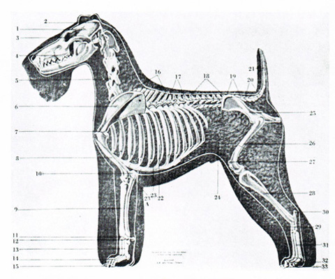
If you are reading the Kerry Blue Terrier Breed Handbook, we will take it for granted that you either have a Kerry or intend to have one. You may even have a "bug" of wishing to show and breed Kerries. Whatever your interest, the more knowledgeable you are about your Kerry, the more you will enjoy him or her. The Kerry skeleton is made up of the same bones as we have, with the same names and the same uses, except that Kerries walk on four legs. The skeleton is the foundation; it forms a cage to protect vital organs and a framework for the flesh (muscles, tendons, etc.), the skin and coat. You may acquaint yourself with the anatomy of your Kerry by using the following explanations, numbered to correspond to the numbers on the illustration, and by feeling the structure of your dog. Head 1 – SKULL is the bony framework which houses the brain, sensory organs (eyes, ears, etc.) It extends from above the eyes to the OCCIPUT (2) the high point at the back of the head. 3 - ZYGOMATIC ARCH - cheek bone – it can be felt on the sides of the skull, and joins the skull to the muzzle by forming the back part of the upper jaw. It plays an important part in determining the size and shape of the head. 4 - Muzzle – foreface – is the portion of the head in front of the eyes. It is made up of the upper and lower jaws, teeth, nose, etc. 5- Neck- seven cervical vertebrae – the first two, immediately back of the occiput, differ in shape from the remaining five, and it is the manner in which they are joined that governs the arch of the neck. Forequarters 6 - SCAPULA – shulderblade – a flat bone, roughly triangular, bearing a conspicuous spine on the lateral aspect. The spine of scapula extends down the middle and terminates distally in the ACROMION PROCESS (7). The. angle of the layback is measured by using the spine as one side of the angle. 8 – HUMERUS – upper arm – between the shoulder joint and elbow joint. 9 – RADIUS AND ULNA- lower arm – the RADIUS is the shorter of the two bones. The ULNA is longer, it extends above the elbow joint and behind the HUMERUS (upper arm) to form the OLECRANON PROCESS (10), tip of the elbow – and is so constructed to permit only forward movement of the forearm. 11 – CARPAL BONES – wrist or pastern joint – comprises seven small bones, arranged in two rows. The ACCESSORY or PISIFORM CARPAL (12) is a short rod of the bone projecting slightly at the back of the joint. Muscles attached to it provide the leverage of the point and bones below it. 13- METACARPAL BONES – hand or pastern bones – there are five in the pastern of the dog. 14- PHALANGES – fingers – there are five or these bones, four of witch form the paw of the dog. The fifth, or thumb, is the dewclaw, which for the comfort of dog should be removed at an early age. 15- TOENAILS – each of the four paw bones has a toenail, as does the dewclaw. Function – There are no bony joints connecting the FOREQUARTER ASSEMBLY to the body, which is achieved by a complex network of powerful muscles. If the SCAPULA (shoulder) is set on the body at an approximate angle of 45? to the perpendicular, and if it is approximately the same length as the HUMERUS (upper arm) at an approximate angle of 90? at the shoulder joint, and 135? at the elbow joint – then a perpendicular line dropped from the tip of the shoulder should touch the OLECRANON (tip elbow), the ACCESSORY or PISIFORM CARPAL (wrist joint), and tough the ground about ? "- 1" behind the paw (the pastern should have a slight slope to cushion front action). Thus your Kerry will have maximum reach forward, and follow through back, lifting his paws no more than 2"-3" of the ground. Body A- SPINAL COLUMN – top line and tail your Kerry 16-THORACIC VERTEBRAE – withers. (There are several versions as to the interpretation if the world. For our purposes it is meant to cover the first 9 vertebrae.) The top spines of these vertebrae slope toward the tail. If you follow down the flatter neck vertebrae with your hand you can easily feel the change in bone formation. There is a slight rise to the third or fourth vertebra approximately above the tip of the shoulder, and then the vertebrae slope gently towards the rear. 17 - THORACIC VERTEBRAE – back, or saddle – last four the bone formation changes again, the spines on these vertebrae are shorter. They form a reasonably flat portion of the dog's top line. 18 – LUMBAR VERTEBRAE – loin- there is a very marked change in the stronger, and have a slightly convex curve, which rises and falls to cushion the rear gait. 19 - SACRUM – 20- COCCYEAL VERTEBRAE – croup assembly – the SACRUM consists of three fused, roughly triangular shaped. The COCCYGEAL VERTEBRAE are the three to four tail bones within the body. 21- EXTERNAL COCCYGEAL VERTEBRAE – tail – the number of these bones depends on docking. Function – This assembly is held together by a network of powerful muscles within the body. It consists of a series of convex and concave curves as does the human spine, and for the same reason: to cushion the shock of walking or gaiting. It terminates in the CROUP ASSEMBLY witch slopes toward the external tail at an angle of approximately 15 to the horizontal. The length and slope of the croup governs the tail set. B- THORACIC CAVITY – chest – is the space enclosed by the rib cage, the thirteen thoracic vertebrae, and the sternum containing the heart and lungs. 22 - RIBS – there are thirteen pairs of ribs. The first nine pair attach to the sternum. The next three pair attach to the sternum by cartilage. The thirteenth pair are "floating" ribs. 23 - STERNUM – breast bone or brisket – forms the floor of the chest. It is roughly parallel to the grounds forward to the elbow joints, the curves up and forward to appear of the shoulder joints, forming the TIP of the STERNUM (23A). Function - the ribs are joined to the vertebrae by joints, and curve out from it at approximately one level, also forming a convex curve to the rear at approximately 45? to the spine. It is the outward curve that determines the spring of rib. The ribs under the shoulder and behind the elbow are fairly flat to permit a free and at the same time close movement of the elbow joint. The ribs the widen toward the rear. The rib cage must have sufficient width and depth to adequately house the heart and lungs, and be held together by pliable muscles to ensure ease of breathing. If the rib cage is correctly formed, the forequarters will be able to fit well back on it, the tip of the STERNUM approximately 1" ahead of the shoulder joint. The FOREQUARTER ASSEMLY will then be in the correct position to ensure a true gait when you Kerry move. C- ABDOMEN – contains the intestines, and is the portion of the body behind the diaphragm (a large muscular tissue) which separate them from the heart and lungs. 24 – FLANK- tuck up – is the side of the body between the last rib and pelvis (hip). Hindquarters 25 – OS COXIAE – pelvis or hipbones – ileum, isocheim and pubes – the former two bones form the upright wings we can feel, and join to form the acetabulum, or the socket into which the upper end of the femur (upper thigh) fits, to form a ball and socket joint, The foremost tip is felt along the spine at the croup, and the lower tip below the tail (pin or seat bone). The isocheim also helps form the floor of the pelvis with the pubic bones, creating an aperture though which the internal organs make contact with the external; hence the size, shape and placement of these bones is of the utmost importance in your brood bitch. 26 – FEMUR – Upper thigh – the largest bone in the body. 27- PATELLA – knee cap – is held in place with strong tendons over the stifle (knee) joint which is between the upper and lower thighs. 28 - TIBIA and FIBULA – lower thigh – TIBIA is the shin bone. The FIBULA is the slenderest bone in the body. 29 - TARSAL BONES – hock or ankle joint. The most important of the seven bones is the OS CALCIS – (30) tip of hock. It is this bone to which the very powerful muscles of the hindquarters are attached by means of the easily felt Achilles tendon. 31 – METATARSAL BONES – foot, hock or rear pastern – resembles the front pastern and also has five bones. 32 PHALANGES – toes – similar to the front paw, except the fifth toe or dewclaw is rarely present and is removed if it does appear. 33 NAILS – similar to the nails on front paws. Function – The HINDQUARTER ASSEMBLE is joined to the body by means of a practically rigid gristly attachment or joint between the ileum and the SACRUM. If the bone lengths of this assembly are in correct proportion to each other, the pelvis approximately the same length as the FEMUR (upper thigh), the TIBIA and FIBULA (lower thigh) longer, and METATARSALS (rear pastern) short and if the resultant angles are approximately hip joint 90? , stifle joint 90?, hock joint 125? , rear pastern 90? to ground – then a perpendicular line dropped from lower tip of isocheim (pin bone) will fall just at the front of the paw of a Kerry at normal stance. Gait If the bone lengths and angels of the FOREQUARTER and HINDQUARTER ASSEMBLIES are correct and balance each other, and if these ASSEMBLIES are correctly placed on the body (as explained) – then you have the foundation for correct movement. BUT –the skeleton is coordinated by pliable elastic muscles, tendons, etc., which are governed by impulses form the brain. Therefore GAIT is dependent on temperament, condition and environment as well as the skeletal structure. Continuous research is being done on the Kerry, and we hope we whetted your curiosity sufficiently so that you will wish to delve more deeply in this intricate but fascinating subject.
| ||